Laboratory for Surface Diagnostics
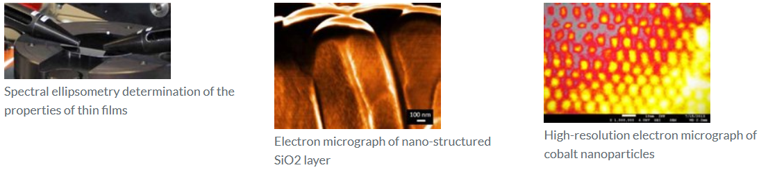
The properties of materials and the interaction of materials with the environment are primarily determined by surface conditions. By using plasma technology it is possible to specifically modify almost any surface property and to generate new materials with special functions that way. The analysis of surfaces is one of the special fields of the INP. The existing spectrum of equipments, the knowledge for operation and the methods for analysing measurement data are continuously extended and improved. Thus, it has been possible for INP to produce highly precise results all the times and to position itself in the world-wide top-level research for years. The production of precise results and the positioning in the international top-level research are guiding principles of the institute.
Determination of the chemical composition and bindings in the surface
- High-resolution X-ray photoelectron spectroscopy (XPS)
- Lateral resolution >27 µm
- Energy resolution: 1 eV
- 2D imaging mode
- Ion source (argon or coronene C24H12) for the cleaning of the surface and production of depth profiles
- FT infrared spectroscopy
- Qualitative chemical analysis of functional groups in MIR spectral range
- Sample specific configurations: ATR, IRRAS, transmission
- FTIR mapping of planar samples (ATR microscopy)
Determination of the morphology of the surface
- Atomic force microscopy (AFM)
- Scan range:
- max. 100 µm x 100 µm for overview images
- ≤ 10 µm x 10 µm for detailed images
- ≤ 1 µm x 1 µm for high-resolution images
- Different measurement modes:
- C-AFM static scanning in the contact mode
- NC-AFM oscillating scanning in non-contact mode
- IC-AFM oscillating scanning in approximated mode
- LFM scanning considering lateral forces (friction measurement)
- Maximum measurable heigth difference: 6 µm
- Scan range:
- Profilometry
- Height resolution: 6.5 µm, 65 µm, 131 µm, 2 mm
- Maximum sample size: 165 mm x 165 mm x 45 mm
Maximum lateral resolution: 4 nm
- Optical microscope with 3D function
- Reflected light and transmitted light microscopy
- Resolutions: 25x, 50x, 200x, 500x
- Stereoscopic images (3D)
Including digital image recording, video recording
Determination of the transmission/reflection of the surface
- UV-Vis spectral photometry
- Wavelength range: 200 nm to 1000 nm
- Optical constants (refraction index, extinction coefficient) and geometric layer thickness of single layers
- Estimation of the band gap of semiconducting materials
Determination of the adhesive strength of the coating
- Taber test
- Characterization of the scratch and abrasion resistance of planar samples
- Calotest (calotte grinding)
- Layer thickness measurement from 200 nm
- Diagnostics of multilayer structures
- Characterization of the abrasion parameters of the coatings
- Ultrasonic bath
- Frequency: 35 kHz
- Power: 2 x 35 W
- Different test liquids
Determination of the contact angle/surface energy
- Contact angle measuring instruments
- Minimum drop volume: 0.5 µl
- Testing with up to 4 liquids
- Water
- Ethylene glycol
- Diiodomethane
- Optional 4th liquid
- Including video function