16th National Health Industry Conference 2021
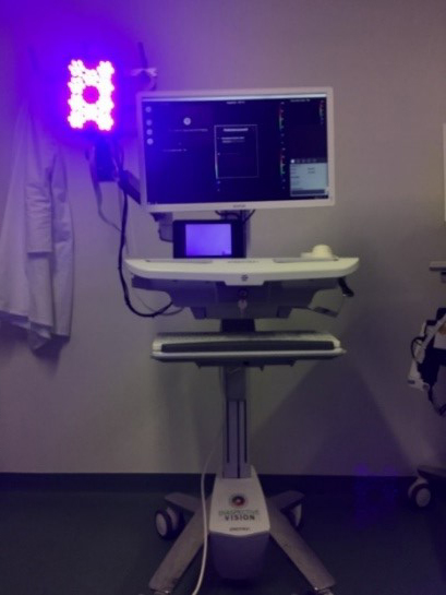
In a joint project funded by TBI (Technology Consulting Institute) with Diaspective Vision GmbH and scientific partners (Wismar University of Applied Sciences, Greifswald University Medical Centre), the idea of making non-invasive wound germs visible using a special camera was successfully implemented. The hyperspectral camera developed by Diaspective Vision GmbH for this purpose was extended to a wavelength range. This enabled the detection of bacteria using autofluorescence.
In the interests of wound patients, the expansion of this innovative optical diagnostic method in further research projects is now on the agenda. To this end, local infection sites will be correlated with physiological parameters such as oxygen saturation in clinical tests and the bacterial load in patients will be measured. The scientists' goal is to find out whether an infection or natural bacterial colonisation is present. This is important in order to decide whether antibiotic therapy is necessary for wound healing.
In addition, the researchers are pursuing the vision of identifying infectious agents directly in the wound so that broad-spectrum antibiotics can be avoided in therapy in the long term to minimise the development of resistance.
Preclinical and clinical applications of hyperspectral diagnostics were carried out in advance at the Kompetenzzentrum Diabetes Karlsburg (KDK). For this purpose, both in vitro experiments (in the laboratory) and in vivo experiments in a study with patients with diabetic foot syndrome were carried out. The qualitative bacterial load of chronic wounds was analysed in comparison with standard microbial diagnostics. The tests showed that fluorescence spectroscopy in combination with hyperspectral analysis can detect different bacteria both in laboratory cultures (on agar and meat) and in infected wounds in patients, and can also display them in spatial resolution.